
You can download Nervous System information throughly from here without any difficulty.
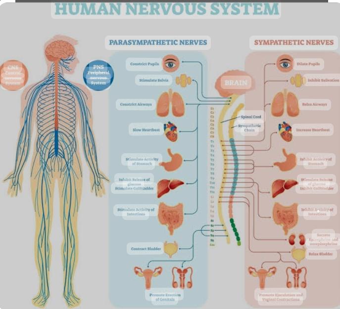
The nervous system consists of the brain, spinal cord, sensory organs, and all of the nerves that connect these organs with the rest of the body. Together, these organs are responsible for the control of the body and communication among its parts. The brain and spinal cord form the control center known as the central nervous system (CNS), where information is evaluated and decisions made. The sensory nerves and sense organs of the peripheral nervous system (PNS) monitor conditions inside and outside of the body and send this information to the CNS. Efferent nerves in the PNS carry signals from the control center to the muscles, glands, and organs to regulate their functions.
Nervous System Anatomy
Nervous Tissue
The majority of the nervous system is tissue made up of two classes of cells: neurons and neuroglia
Neurons.
Neurons, also known as nerve cells, communicate within the body by transmitting electrochemical signals. Neurons look quite different from other cells in the body due to the many long cellular processes that extend from their central cell body. The cell body is the roughly round part of a neuron that contains the nucleus, mitochondria, and most of the cellular organelles. Small tree-like structures called dendrites extend from the cell body to pick up stimuli from the environment, other neurons, or sensory receptor cells. Long transmitting processes called axons extend from the cell body to send signals onward to other neurons or effector cells in the body.
There are 3 basic classes of neurons: afferent neurons, efferent neurons, and interneurons.
1. Afferent neurons. Also known as sensory neurons, afferent neurons transmit sensory signals to the central nervous system from receptors in the body.
_2. ÊEfferent neurons. Also known as motor neurons, efferent neurons transmit signals from the central nervous system to effectors in the body such as muscles and glands._
3. Interneurons. Interneurons form complex networks within the central nervous system to integrate the information received from afferent neurons and to direct the function of the body through efferent neurons.
Neuroglia.
Neuroglia, also known as glial cells, act as the ”helper” cells of the nervous system. Each neuron in the body is surrounded by anywhere from 6 to 60 neuroglia that protect, feed, and insulate the neuron. Because neurons are extremely specialized cells that are essential to body function and almost never reproduce, neuroglia are vital to maintaining a functional nervous system.
Brain The brain, a soft, wrinkled organ that weighs about 3 pounds, is located inside the cranial cavity, where the bones of the skull surround and protect it. The approximately 100 billion neurons of the brain form the main control center of the body. The brain and spinal cord together form the central nervous system (CNS), where information is processed and responses originate. The brain, the seat of higher mental functions such as consciousness, memory, planning, and voluntary actions, also controls lower body functions such as the maintenance of respiration, heart rate, blood pressure, and digestion.
Spinal Cord The spinal cord is a long, thin mass of bundled neurons that carries information through the vertebral cavity of the spine beginning at the medulla oblongata of the brain on its superior end and continuing inferiorly to the lumbar region of the spine. In the lumbar region, the spinal cord separates into a bundle of individual nerves called the cauda equina (due to its resemblance to a horse’s tail) that continues inferiorly to the sacrum and coccyx. The white matter of the spinal cord functions as the main conduit of nerve signals to the body from the brain. The grey matter of the spinal cord integrates reflexes to stimuli.
Nerves Nerves are bundles of axons in the peripheral nervous system (PNS) that act as information highways to carry signals between the brain and spinal cord and the rest of the body. Each axon is wrapped in a connective tissue sheath called the endoneurium. Individual axons of the nerve are bundled into groups of axons called fascicles, wrapped in a sheath of connective tissue called the perineurium. Finally, many fascicles are wrapped together in another layer of connective tissue called the epineurium to form a whole nerve. The wrapping of nerves with connective tissue helps to protect the axons and to increase the speed of their communication within the body.
Afferent, Efferent, and Mixed Nerves. Some of the nerves in the body are specialized for carrying information in only one direction, similar to a one-way street. Nerves that carry information from sensory receptors to the central nervous system only are called afferent nerves. Other neurons, known as efferent nerves, carry signals only from the central nervous system to effectors such as muscles and glands. Finally, some nerves are mixed nerves that contain both afferent and efferent axons. Mixed nerves function like 2-way streets where afferent axons act as lanes heading toward the central nervous system and efferent axons act as lanes heading away from the central nervous system.
Cranial Nerves. Extending from the inferior side of the brain are 12 pairs of cranial nerves. Each cranial nerve pair is identified by a Roman numeral 1 to 12 based upon its location along the anterior-posterior axis of the brain. Each nerve also has a descriptive name (e.g. olfactory, optic, etc.) that identifies its function or location. The cranial nerves provide a direct connection to the brain for the special sense organs, muscles of the head, neck, and shoulders, the heart, and the GI tract.
Spinal Nerves. Extending from the left and right sides of the spinal cord are 31 pairs of spinal nerves. The spinal nerves are mixed nerves that carry both sensory and motor signals between the spinal cord and specific regions of the body. The 31 spinal nerves are split into 5 groups named for the 5 regions of the vertebral column. Thus, there are 8 pairs of cervical nerves, 12 pairs of thoracic nerves, 5 pairs of lumbar nerves, 5 pairs of sacral nerves, and 1 pair of coccygeal nerves. Each spinal nerve exits from the spinal cord through the intervertebral foramen between a pair of vertebrae or between the C1 vertebra and the occipital bone of the skull.
Meninges
The meninges are the protective coverings of the central nervous
system (CNS). They consist of three layers: the dura mater,
arachnoid mater, and pia mater.
Dura mater.ÊThe dura mater, which means ”tough mother,” is the thickest, toughest, and most superficial layer of meninges. Made of dense irregular connective tissue, it contains many tough collagen fibers and blood vessels. Dura mater protects the CNS from external damage, contains the
cerebrospinal fluid that surrounds the CNS, and provides blood to the nervous tissue of the CNS.
Arachnoid mater. The arachnoid mater, which means ”spider-like mother,” is much thinner and more delicate than the dura mater. It lines the inside of the dura mater and contains many thin fibers that connect it to the underlying pia mater. These fibers cross a fluid-filled space called the subarachnoid space between the arachnoid mater and the pia mater.
Pia mater. The pia mater, which means ”tender mother,” is a thin and delicate layer of tissue that rests on the outside of the brain and spinal cord. Containing many blood vessels that feed the nervous tissue of the CNS, the pia mater penetrates into the valleys of the sulci and fissures of the brain as it covers the entire surface of the CNS.
Cerebrospinal Fluid
The space surrounding the organs of the CNS is filled with a clear fluid known as cerebrospinal fluid (CSF). CSF is formed from blood plasma by special structures called choroid plexuses. The choroid plexuses contain many capillaries lined with epithelial tissue that filters blood plasma and allows the filtered fluid to enter the space around the brain.
Newly created CSF flows through the inside of the brain in hollow spaces called ventricles and through a small cavity in the middle of the spinal cord called the central canal. CSF also flows through the subarachnoid space around the outside of the brain and spinal cord. CSF is constantly produced at the choroid plexuses and is reabsorbed into the bloodstream at structures called arachnoid villi.
Cerebrospinal fluid provides several vital functions to the central nervous system:
1. CSF absorbs shocks between the brain and skull and between the spinal cord and vertebrae. This shock absorption protects the CNS from blows or sudden changes in velocity, such as during a car accident.
2. The brain and spinal cord float within the CSF, reducing their apparent weight through buoyancy. The brain is a very large but soft organ that requires a high volume of blood to function effectively. The reduced weight in cerebrospinal fluid allows the blood vessels of the brain to remain open and helps protect the nervous tissue from becoming crushed under its own weight.
3. CSF helps to maintain chemical homeostasis within the central nervous system. It contains ions, nutrients, oxygen, and albumins that support the chemical and osmotic balance of nervous tissue. CSF also removes waste products that form as byproducts of cellular metabolism within nervous tissue.
Sense Organs
_All of the bodies many sense organs are components of the nervous system. What are known as the special senses and vision, taste, smell, hearing, and balance & are all detected by specialized organs such as the eyes, taste buds, and olfactory epithelium. Sensory receptors for the general senses like touch, temperature, and pain are found throughout most of the body. All of the sensory receptors of the body are connected to afferent neurons that carry their sensory information to the CNS to be processed and integrated.